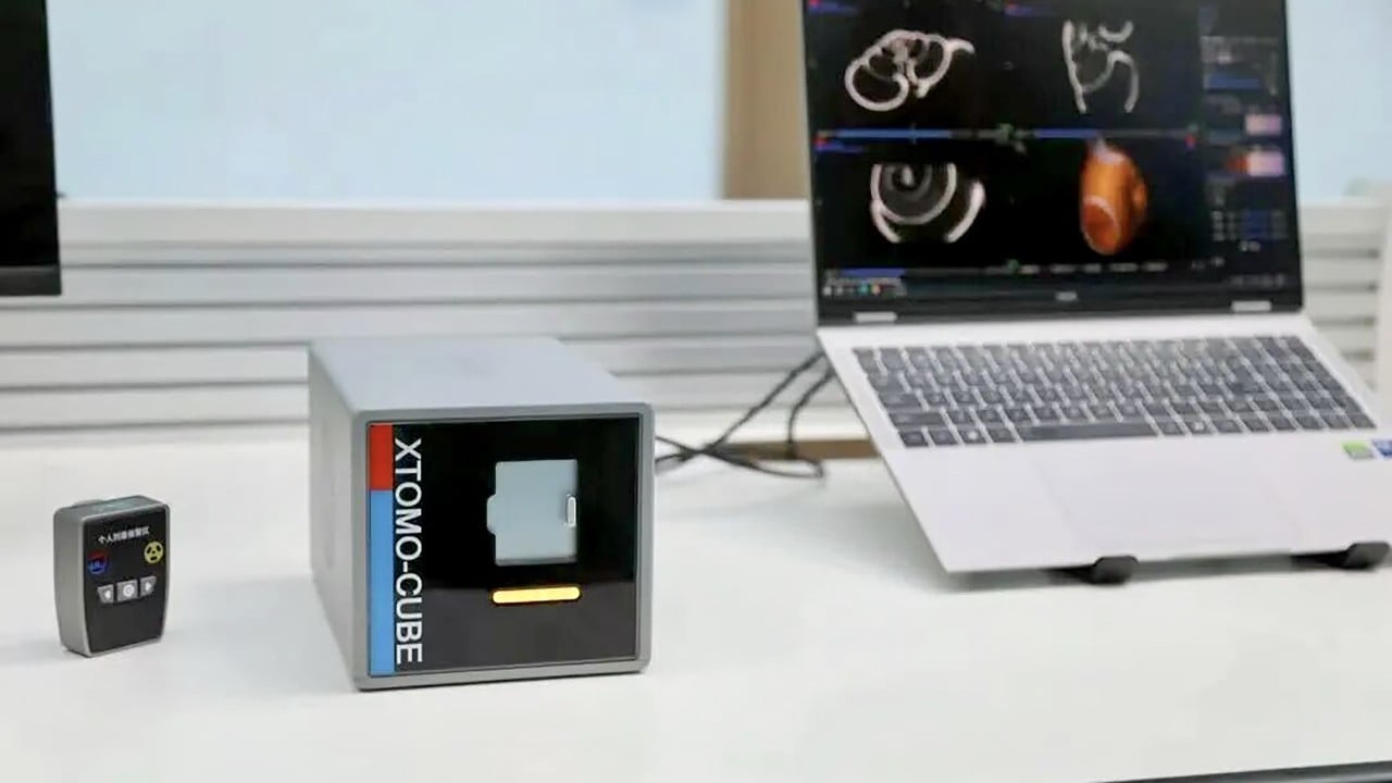
Chinese researchers have built a functional computed tomography scanner weighing just six kilograms, shrinking a multi-tonne hospital fixture into a portable unit that fits inside a standard backpack. For decades, advanced medical imaging required moving patients to stationary, heavy-duty machines housed in reinforced hospital wings. This new six-kilogram device dismantles that entire operational model. Diagnostic capability is no longer chained to concrete floors and high-voltage substation feeds. Instead, high-resolution cross-sectional scans can now be performed in cramped ambulances, backcountry clinics, or disaster zones where minutes determine survival.
The Weight of Tradition
To understand why a six-kilogram computed tomography unit represents a structural shift in medicine, look at the evolution of the hardware itself. Since the invention of computed tomography in the early 1970s, engineers fought a continuous battle against mechanical inertia. A standard hospital scanner is an engineering monument. It requires a massive rotating gantry spinning an X-ray tube and a curved array of detectors at high speeds around a patient aperture. The centrifugal forces generated by this high-speed rotation demand heavy steel frames, thick tungsten shielding to contain scatter radiation, and dedicated cooling systems to prevent thermal meltdown.
These machines regularly tip the scales at two thousand kilograms or more. They demand reinforced flooring, specialized electrical wiring capable of drawing massive bursts of current, and climate-controlled environments. Hospitals treated these installations as permanent real estate investments. Bringing a patient to the machine was non-negotiable because the machine could never go to the patient.
That immobility created a silent bottleneck in emergency medicine. A stroke victim sitting in a rural clinic fifty miles from a regional medical center loses precious brain tissue during every minute of transit. A trauma patient bleeding internally inside a remote mountain pass cannot wait for a forty-minute ambulance ride to the nearest tertiary hospital just to confirm the site of arterial rupture. The medical device industry accepted this limitation as a law of physics. Rotating heavy hardware requires mass, and mass requires stability.
Engineering the Miniature
Bending that law required a complete reinvention of how X-ray projection and image reconstruction operate. Traditional scanners rely on mechanical rotation. The gantry spins continuously to capture angular projections from every direction. Scaling that architecture down to six kilograms means eliminating the massive moving gantry entirely or reimagining its mechanical footprint.
While specific proprietary architectural details from the Chinese research team continue to circulate primarily within specialized engineering journals and domestic patent filings, the underlying physics of portable computed tomography point toward solid-state emitter arrays and stationary gantry designs. Instead of spinning a physical X-ray tube around the patient, engineers increasingly rely on distributed carbon-nanotube field-emission X-ray sources. These cold-cathode tubes emit X-rays electronically without heating filaments or requiring mechanical rotation. Multiple emitters are arranged in a stationary ring, firing sequentially in milliseconds.
This electronic switching replaces physical motion with digital sequencing. Without heavy motors, slip rings, bearings, and heavy balancing weights, the physical mass of the gantry collapses. The detectors themselves have also undergone radical downsizing. Modern high-density scintillation crystals coupled with advanced silicon photomultipliers allow for compact, lightweight detector rings that capture high-fidelity photon data without bulky cooling housings.
The Power and Thermal Dilemma
Miniaturizing the hardware is only half the battle. Power supply and thermal management present severe engineering barriers that have historically killed portable imaging projects.
Generating diagnostic-quality X-rays requires high voltage and substantial electrical current. A standard hospital scanner taps directly into heavy-duty commercial power grids, pulling instantaneous power loads that would melt standard residential wiring. A six-kilogram portable unit must operate on battery power or standard auxiliary vehicle outlets without sacrificing beam penetration or image clarity.
To achieve this, researchers optimized low-dose X-ray protocols alongside advanced iterative reconstruction algorithms. By relying on computational math rather than brute-force radiation intensity, the system can produce diagnostic images using a fraction of the photon output. Less radiation means lower power consumption, which in turn shrinks the battery pack requirements down to manageable proportions.
Thermal management represents an equally formidable hurdle. When X-ray tubes fire, they generate immense heat. In large hospital units, liquid cooling loops and heavy heat exchangers dissipate this thermal energy. A backpack-sized device has virtually no room for bulky radiators or fluid pumps. Engineers solved this by utilizing high-efficiency solid-state thermal sinks and intermittent burst-firing schedules designed to dissipate heat naturally between rapid scan cycles.
Clinical Realities in the Field
Hardware specs mean nothing if the clinical output is unusable junk. Radiologists rely on millimeter-level spatial resolution to detect subtle ischemic strokes, hairline fractures, or tiny pulmonary embolisms. Early attempts at portable imaging often produced grainy, artifact-heavy scans that served as little more than educated guesses.
The six-kilogram device bridges this gap through deep-learning image reconstruction. When physical hardware is constrained by weight limits, software must compensate for physical shortcomings. Advanced neural networks trained on millions of high-dose computed tomography scans fill in the gaps left by sparse photon counts and lightweight detector configurations. These algorithms suppress electronic noise, correct for beam-hardening artifacts, and sharpen tissue boundaries in real time.
Consider a hypothetical scenario in a high-altitude rescue operation. A climber suffers a severe head injury on an isolated ridge. Traditionally, first responders must administer basic first aid and organize a hazardous, multi-hour evacuation down treacherous terrain, operating completely blind regarding potential intracranial hemorrhage. With a six-kilogram diagnostic unit carried directly to the tent, a paramedic performs an immediate head scan. The onboard processing unit transmits compressed digital imaging files via satellite uplink to a trauma specialist stationed at a major metropolitan hospital. Within minutes, the medical team confirms a subdural hematoma, altering the evacuation protocol and preparing the surgical theatre before the patient even touches tarmac.
This capability transforms the ambulance from a mobile transport vehicle into an active mobile emergency room. Stroke protocols, which rely entirely on the critical window between symptom onset and drug administration, can now begin inside the residential driveway or the workplace breakroom rather than forty-five minutes later upon arrival at the emergency department.
Market Inertia and Regulatory Hurdles
Despite the engineering triumph, commercializing a six-kilogram computed tomography scanner faces immense friction from entrenched medical hierarchies and regulatory conservatism.
Hospitals operate on capital-intensive procurement cycles. Multi-million-dollar imaging suites are cash cows for major healthcare networks, and institutional buyers are inherently suspicious of disruptive hardware that challenges established workflows. Furthermore, medical regulatory bodies across North America, Europe, and Asia maintain rigorous, slow-moving approval processes for diagnostic imaging devices. Proving that a six-kilogram device matches the diagnostic accuracy of a two-tonne stationary machine requires exhaustive clinical trials, multi-center validation studies, and rigorous safety audits regarding cumulative radiation exposure for field operators.
There is also the operator training problem. Operating a standard hospital scanner requires specialized radiological technologists who spend years mastering patient positioning, protocol selection, and radiation safety. Deploying a portable unit into the hands of general paramedics, military medics, or rural nurses requires foolproof software interfaces, automated positioning guides, and fail-safe radiation containment protocols. If an untrained operator scatters X-rays inside an unshielded ambulance or misinterprets an image artifact, the liability implications for the manufacturer are catastrophic.
The Global Manufacturing Shift
The emergence of this technology highlights a broader shift in global medical device manufacturing. For decades, Western conglomerates dominated the high-end diagnostic imaging market, controlling patents for superconductive magnets, high-speed gantries, and precision X-ray tubes. Companies based in Germany, the Netherlands, and the United States set the technological benchmarks and commanded premium pricing across global healthcare systems.
By successfully miniaturizing complex radiological equipment, Chinese researchers and domestic manufacturers are signaling a strategic pivot from imitation to fundamental innovation. This is not merely an incremental upgrade to an existing product line; it is the creation of an entirely new category of medical hardware. Domestic healthcare infrastructure in China, characterized by vast rural populations separated from top-tier urban medical centers, provides a massive testing ground and immediate internal market for portable diagnostic tools.
When a domestic manufacturer can supply every remote county clinic, frontline military unit, and emergency response vehicle with lightweight computed tomography capability, the economic power balance shifts. Western medical equipment manufacturers can no longer rely on the assumption that high-end imaging requires heavy, expensive infrastructure. They are now forced to respond to a standard of mobility that redefines patient care expectations worldwide.
The Final Diagnostic Frontier
The boundary between the hospital and the outside world is dissolving. As power densities improve, battery technologies advance, and artificial intelligence continues to refine low-dose image processing, diagnostic tools will continue to shrink. The six-kilogram computed tomography scanner is not an isolated laboratory curiosity. It represents the opening salvo in a wholesale decentralization of advanced medicine.
When heavy diagnostic machinery can be packed into a backpack and carried into the most hostile environments on earth, the traditional architecture of healthcare delivery becomes obsolete. Patients will no longer travel to the machine. The machine will travel to the patient, wherever they happen to be, shattering the limits of time and geography that have constrained emergency medicine for over a century. The hardware has crossed the threshold from immovable monument to mobile instrument, leaving traditional medical infrastructure scrambling to catch up.