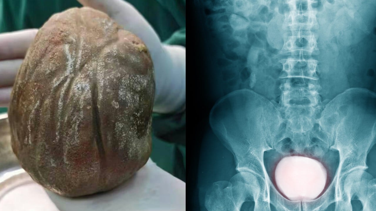
The formation of a 1.3-kilogram urolithiasis within a human bladder represents a catastrophic failure of urinary homeostasis. While mainstream reporting treats such cases as medical anomalies or sensational curiosities, an analytical breakdown reveals them as predictable outcomes of prolonged, unmitigated mechanical and chemical imbalances. To understand how a calculus reaches the mass of a standard laptop computer inside a living organ, one must examine the specific intersection of urinary stasis, bacterial enzymatic activity, and patient-side diagnostic neglect.
The progression from micro-crystalline nucleation to a 1.3kg obstructive mass follows a strict thermodynamic and mechanical trajectory. By analyzing the structural pillars of giant bladder stone development, clinicians and analysts can map the specific physiological compounding loops that allow these pathologies to accelerate over a multi-year timeline.
The Tri-Focal Etiology of Giant Calculi
The development of an extreme bladder stone relies on three interdependent physiological vectors. If any of these vectors is interrupted, stone growth plateaus. When all three operate concurrently over a multi-year timeline, growth accelerates exponentially.
+------------------------+
| Urinary Stasis         | -> Bladder outlet obstruction prevents crystal clearance
+------------------------+
            |
            v
+------------------------+
| Chronic Infection      | -> Urease-producing bacteria elevate pH levels
+------------------------+
            |
            v
+------------------------+
| Substrate Abundance   | -> Hypercalciuria and hyperoxaluria fuel crystal growth
+------------------------+
1. The Stasis Vector (Mechanical Obstruction)
Urinary stasis is the foundational requirement for extreme calculus retention. In a healthy urological ecosystem, micro-crystals of calcium oxalate or uric acid form regularly but are flushed during normal micturition before aggregation occurs. For a stone to achieve a mass measured in kilograms, an anatomical or functional bottleneck must exist.
The primary driver is typically a bladder outlet obstruction (BOO). In aging male populations, this stems from benign prostatic hyperplasia (BPH) or urethral strictures. When the bladder cannot empty completely, a residual volume of urine remains stagnant. This residual pool serves as an incubator, allowing crystals to settle, adhere to the urothelium, and form a permanent nidus.
2. The Biochemical Environment (The pH Shift)
The chemical composition of the stone dictates its growth rate. While calcium oxalate stones grow slowly via metabolic supersaturation, giant stones—particularly those exceeding 500 grams—are frequently composed of struvite (magnesium ammonium phosphate) or a combination of calcium phosphate and uric acid.
The acceleration loop begins with chronic, low-grade urinary tract infections (UTIs) caused by urease-producing bacteria such as Proteus mirabilis, Klebsiella pneumoniae, or Pseudomonas.
$$\text{CO(NH}_2)_2 + \text{H}_2\text{O} \xrightarrow{\text{Urease}} 2\text{NH}_3 + \text{CO}_2$$
$$\text{NH}_3 + \text{H}_2\text{O} \rightleftharpoons \text{NH}_4^+ + \text{OH}^-$$
The generation of free hydroxyl ions ($\text{OH}^-$) drives the urinary pH above 7.2. This alkaline shift drastically reduces the solubility of phosphate ions, precipitating large quantities of struvite and carbonatapatite out of solution and onto the existing stone matrix.
3. Patient Psychodynamics and Habituation
The third, often overlooked pillar is the neurological and behavioral habituation of the patient. A stone does not reach 1.3 kilograms in months; it requires years—in this specific index case, three years of continuous growth. The human bladder adapts to slow, progressive volume displacement.
As the stone displaces urine capacity, the detrusor muscle undergoes compensatory hypertrophy. The patient experiences increased frequency, urgency, and dysuria, but because the onset is gradual, a process of sensory habituation occurs. The patient normalizes severe pain and alters their lifestyle to accommodate the dysfunction, delaying clinical intervention until total mechanical obstruction occurs.
The Growth Function and Mass Acceleration
The kinetic growth of a bladder stone is not linear. It follows a surface-area-dependent acceleration model. The rate of mass accumulation ($\frac{dM}{dt}$) is directly proportional to the available surface area ($A$) of the calculus exposed to supersaturated urine.
$$M \propto V \implies M = k \cdot r^3 \quad (\text{where } k = \frac{4}{3}\pi\rho)$$
$$A = 4\pi r^2 = 4\pi \left(\frac{M}{k}\right)^{2/3}$$
As the radius ($r$) increases, the total surface area expands, providing more binding sites for crystal accretion.
Early-stage growth (Years 1–2) is characterized by a slow mass increase as the stone establishes its core matrix.
Late-stage growth (Year 3) features rapid mass acceleration. The stone acts as a massive chemical sink, drawing ions out of the passing urine at an accelerated rate. This explains how a patient can tolerate a growing stone for years with mild to moderate symptoms, only to rapidly crash into acute renal failure when the stone fills the entire vesical vault.
Secondary Systemic Failures: The Cascading Pathology
A giant bladder stone does not exist in isolation; it alters the entire upper and lower urinary tract architecture. The physical presence of a 1.3kg mass causes predictable secondary pathologies.
Bilateral Hydronephrosis and Nephron Atrophy
The stone eventually occupies the entire bladder lumen, compressing the ureterovesical junctions (UVJs). This creates a functional bilateral ureteral obstruction. Urine cannot exit the ureters, causing backward hydrostatic pressure to build through the renal pelvis and into the nephrons.
The resulting hydronephrosis dilates the renal calyces, compresses the renal parenchyma, and disrupts the glomerular filtration rate (GFR). Prolonged exposure to this elevated backpressure causes irreversible nephron loss and chronic kidney disease (CKD).
Detrusor Muscle Remodeling and Fibrosis
To overcome the massive mechanical resistance of the stone, the bladder wall must generate extreme voiding pressures. This results in severe trabeculation and hypertrophy of the detrusor muscle. Over time, chronic ischemia and mechanical strain cause the muscular tissue to be replaced by collagen deposits. The bladder loses its elasticity (compliance), converting the organ from a low-pressure reservoir into a high-pressure, non-functional fibrotic sac. This damage is often permanent, persisting even after the stone is surgically removed.
Surgical Extraction Architecture: Risks and Execution
The extraction of a 1.3kg bladder stone rules out minimally invasive urological techniques. Endoscopic lithotripsy (laser or electrohydraulic) is mathematically non-viable; the energy required to fragment a stone of this density and volume would cause catastrophic thermal and mechanical injury to the bladder wall, and the procedure would require dozens of operative hours.
The definitive intervention is an open suprapubic cystolithotomy. This procedure presents distinct surgical challenges:
- Incision Dynamics: The cystotomy incision must be large enough to deliver the stone without tearing the heavily vascularized, hypertrophied bladder wall.
- Vascular Management: The bladder wall in these patients is highly hyperemic and prone to severe hemorrhage upon incision.
- Post-Extraction Voiding Dysfunction: Removing the stone leaves a massive void. The overstretched detrusor muscle is frequently acontractile post-surgery, requiring prolonged suprapubic or transurethral catheterization to allow the bladder wall to remodel and regain tone.
Diagnostic Frameworks for High-Risk Populations
To prevent cases from escalating to this extreme magnitude, clinical protocols must identify the early markers of urinary stasis and crystalline aggregation. Relying solely on patient-reported pain is an unreliable strategy due to individual differences in pain tolerance and sensory habituation.
| Diagnostic Modality | Targeted Biomarker / Pathology | Clinical Action Threshold |
|---|---|---|
| Point-of-Care Ultrasound (POCUS) | Post-void residual (PVR) volume, acoustic shadowing | PVR > 100 mL indicates structural or functional stasis requiring investigation. |
| Urinalysis & Microscopy | pH trends, persistent microhematuria, triple phosphate crystals | Persistent pH > 7.0 demands a urine culture for urease-producing pathogens. |
| KUB Radiography | Radiopaque pelvic masses, macro-calcifications | Any visible density in the lesser pelvis requires immediate tomographic sizing. |
Implementing routine post-void residual tracking via ultrasound in patients presenting with recurrent UTIs or progressive lower urinary tract symptoms (LUTS) cuts off the development loop. If urinary stasis is caught and corrected—whether through pharmaceutical management of prostate volume or surgical correction of strictures—the environment required for giant calculus synthesis cannot stabilize.