The evolutionary divergence between Homo neanderthalensis and Homo sapiens has traditionally been mapped through the morphometric analysis of adult skeletal structures. However, understanding the precise operational parameters of these differences requires an examination of ontogeny—the developmental trajectory from gestation through adulthood.
A foundational question in paleoanthropology centers on whether the robust skeletal architecture of adult Neanderthals was the product of a accelerated prenatal growth blueprint, or a divergence that materialized post-natally.
Data published in Royal Society Open Science by an international research collective isolates the structural characteristics of Neanderthal development using high-resolution virtual microanatomy. By deploying non-invasive micro-computed tomography (micro-CT) on exceptionally rare infant remains from the Sesselfelsgrotte rock shelter in Lower Bavaria, Germany, researchers have mapped the cellular architecture of ancient development.
The findings establish a critical baseline: prehistoric fetal development shared a highly conserved structural blueprint with modern humans during late-stage gestation, with morphological divergence occurring as a localized, post-natal acceleration driven by environmental and metabolic pressures.
The Microanatomical Architecture of Gestation
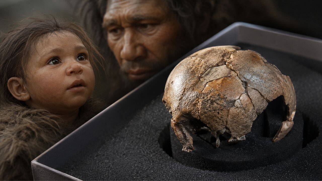
The primary specimen under analysis, designated Sesselfelsgrotte 1, consists of 12 fragmentary bone elements originating from a single late-term perinate or unborn fetus dating to approximately 50,000 to 75,000 years ago. Historically, assessing the developmental age of such fragmented material required destructive histological sectioning. The application of micro-CT bypasses this limitation, yielding three-dimensional reconstructions of internal bone microstructure at a micron scale.
Virtual microanatomy reveals that the bone tissue patterns of Sesselfelsgrotte 1 correspond directly with the standard indicators of a rapidly developing fetal skeleton in the final trimester of pregnancy, specifically matching modern human reference models at 30 to 36 weeks of gestation. The diagnostic features include:
- High Vascularity: A dense network of micro-channels designed to accommodate rapid blood flow and nutrient delivery, characteristic of active osteogenesis.
- Immature Bone Matrix: High concentrations of woven bone, a transient, mechanically weak tissue that serves as the immediate precursor to structured lamellar bone.
- Accelerated Osteogenic Volumetric Rates: Spatial distribution of bone mineralization indicating rapid, un-reformed deposition typical of late-stage fetal growth.
While the overarching developmental trajectory mirrors that of Homo sapiens, structural variations appear when analyzing the skeleton by distinct anatomical zones. The long bones—specifically the femur and humerus—exhibit an advanced degree of cortical compactness and structural organization relative to the cranial vault and jaw fragments. This regional variance indicates that while the global prenatal blueprint remains conserved across both human lineages, Neanderthals possessed an accelerated post-cranial mineralization program that was already active in the womb.
The Deciduous Matrix and Metabolic Stress Factors
In parallel with the fetal skeletal fragments, the team analyzed two deciduous molars (Sesselfelsgrotte 2 and 3) from separate juvenile individuals. Because tooth enamel and dentine form chronologically and do not undergo remodeling like bone tissue, they act as permanent, incremental biological archives of early-life physiology.
Micro-CT imaging of these teeth revealed pronounced mineralization defects embedded deep within the dentine. These structural anomalies are classified as interglobular dentine (IGD). IGD occurs when the unmineralized organic matrix fails to coalesce into a homogeneous calcified structure, leaving distinct, uncalcified fluid-filled gaps between mineralized globules.
Dentine Matrix Formation -> [Systemic Metabolic Insult] -> Interrupted Calcification -> Interglobular Dentine (IGD)
The presence of interglobular dentine provides explicit data regarding the systemic physiological stress profiles of young Neanderthals. The biological mechanisms responsible for generating IGD are highly specific, establishing a direct causal chain back to metabolic disruptions during early development. The primary systemic failures capable of arresting dentine calcification include:
- Hypovitaminosis D: Severe vitamin D deficiency, which fundamentally disrupts the body's homeostatic feedback loops governing calcium management.
- Hypocalcemia: Insufficient dietary calcium intake, causing a critical drop in extracellular fluid calcium concentrations below the threshold required for proper tooth mineralization.
- Impaired Intestinal Absorption: Systemic pathologies or acute gastrointestinal distress that inhibit the active transport of divalent cations across the intestinal epithelium.
Because the development of Neanderthal deciduous molars spans a definitive chronological window—beginning in the third trimester of pregnancy and continuing through approximately the second year of life—these IGD zones serve as an unalterable ledger of physiological hardship. The data demonstrates that ancient populations experienced significant systemic metabolic stress during critical developmental windows, highlighting a shared vulnerability to environmental and nutritional volatility.
Divergence Mechanics in Hominin Ontogeny
Synthesizing the perinate bone microarchitecture with the juvenile dental records allows for the construction of a clear model for Neanderthal ontogeny, separating it into distinct phases of alignment and divergence relative to modern humans.
Phase 1: Late Gestation   --> High structural conservation; overlapping trajectories.
Phase 2: Infancy/Toddler  --> Divergence via somatic acceleration; rapid long-bone elongation.
Phase 3: Late Childhood   --> Convergence of relative growth rates; adult morphologic stabilization.
The initial phase—prenatal growth—is characterized by tight developmental constraints. The metabolic cost of gestation is primarily governed by maternal physiology, which imposes a ceiling on structural divergence. The high degree of overlap in the tissue structures of Sesselfelsgrotte 1 confirms that the foundational biological programming of the genus Homo remained structurally consistent across species lines up until the point of birth.
The secondary phase—infancy and early childhood—marks the operational transition where divergence manifests. Comparative skeletal data from other specimens, such as the 1-to-2-year-old Amud 7 skeleton from Israel, demonstrates that post-natal Neanderthal toddlers grew significantly faster than modern humans. During this stage, somatic growth—particularly the elongation and robustification of the long bones—outpaced dental eruption patterns. This asymmetric development contrasts sharply with Homo sapiens, where dental and skeletal maturation proceed in close, proportionate alignment.
This post-natal developmental shift was an evolutionary requirement for surviving the volatile environments of Pleistocene Eurasia. The accelerated growth of long bones and musculature during infancy minimized the period of extreme juvenile vulnerability. However, this strategy carried a severe metabolic penalty. Supplying the caloric and nutritional energy required to fuel both rapid somatic growth and a large brain created a highly delicate metabolic equilibrium. Any disruption in nutrient availability, such as seasonal food shortages or maternal illness, immediately triggered systemic failures, resulting in the mineralization defects documented in the Sesselfelsgrotte dentine.
Methodological Boundaries and Analytical Uncertainty
A rigorous evaluation of this data requires an explicit acknowledgment of the sample limitations inherent to deep-time paleoanthropology. Worldwide, fewer than ten intact or semi-intact Neanderthal fetuses and newborns have been recovered. Consequently, importing modern human reference standards as a baseline for comparative tissue density remains an analytical necessity, introducing an unavoidable degree of uniformitarian assumption.
Furthermore, while non-invasive micro-CT preserves the physical integrity of highly fragile specimens for future genetic and proteomic sampling, it operates under a structural trade-off. Virtual microanatomy visualizes spatial density variations with high fidelity, but it cannot resolve the finest cellular-level signatures—such as the microscopic lines of von Ebner or individual osteocyte lacunae orientation—that destructive thin-section histology can isolate. Thus, while the presence of IGD is indisputable, pinning down the precise temporal onset or the specific etiology of the metabolic insult remains beyond the reach of imaging technology alone.
The strategic imperative for future research rests on multi-method testing regimes. To maximize the analytical return from these rare fossil assets, institutions must systematically cross-reference virtual microanatomy with micro-sampled stable isotope analysis of carbon, nitrogen, and oxygen from the same teeth. Mapping isotopic shifts directly onto the virtual IGD coordinates will make it possible to isolate the exact environmental or maternal health variables that disrupted early hominin development. This integrated approach will transform isolated anatomical specimens into precise readouts of prehistoric human ecology.